[해외]3D 프린팅이 동맥류 내 미세수술 시뮬레이션을 돕다
- 2020-10-19
- 관리자
○ 본문요약 :
실습 훈련이 거의 없는 외과의에게 수술을 받고 싶어하는 사람은 아무도 없지만, 역사를 통틀어, 적절한 모델이나 시체들을 찾는 것은 경험 많은 의사로 가는 길을 계속 가면서 그들의 절차를 완벽히 하는 많은 조용한 시간의 사치만을 원하는 의대생들에게 도전이었다.
3D 프린팅은 환자, 가족, 의대생, 의사뿐만 아니라 전 세계 의사들을 위한 (수술) 테이블에도 많은 것을 가져다 주었다. 3D 프린팅 모델과 가이드는 신장, 머리와 목 종양, 엉덩이 골절과 같은 장기를 표시하든 보다 나은 진단과 치료를 가능하게 하며 종합적인 시각적 효과를 제공한다. 모든 관련자를 위한 원조
시뮬레이션 기기는 더 복잡하지만 학생들과 의사들에게도 매우 도움이 된다. 이제, 베른 대학의 스위스 의학 연구원들은 최근 출판된 "동맥류 미세 수술 훈련을 위한 신경외과 시뮬레이터 - 신경외과의사와 레지던트가 참여하는 사용자 적합성 연구"에 자세히 설명된 새로운 훈련 방법을 개발했다.
현재 이러한 미세수술에 수반되는 난이도로 인해 두개내 동맥류에 대한 훈련이 제한되어 있다. 의대생과 외과의는 전형적으로 수술 중 관찰과 보조를 통해 경험을 쌓고, 숙련된 의사의 주의 하에 수술을 하며, 비디오를 공부한다. 일부 유형의 가상현실 시뮬레이션을 사용할 수 있지만, 연구자들은 혈액 순환, 유동성 등의 기능까지 포함하는 환자 맞춤형 모델로 기술을 개선하고자 하는 동기를 부여받았지만, 이는 훈련 목적에 매우 중요하지만 역사적으로 부족했던 것이다.
두개내 동맥류 미세수술의 섬세한 성격을 고려할 때, 학생들이 동맥류의 혈관적 특성은 물론, 파열 발생 시 어떤 일이 일어나는지 정확히 이해하는 것이 중요하다. 학생과 의사 훈련은 외과적 과정을 전반적으로 이해해야 하며, 외과적 연관성(막힘)을 알아내는 방법을 배워야 한다. 모델 등 체험교육이 워낙 어려워 훈련된 뇌혈관 신경외과가 계속 부족할 수 있다는 우려다.
본 연구의 목적은 3D 프린터로 제작한 모델을 기반으로 진행형 시뮬레이터를 제작하여 뇌내 동맥류를 클리핑하는 데 있어 주민과 신경외과를 모두 양성할 수 있는 것이었습니다. 사용되는 재료는 다음과 같은 인체 조직을 모방하기 위한 것이다.
-동맥벽파티
-두께
-탄력성
-맥동혈류
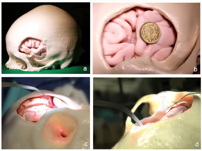
훈련 연구 중 모델 표현: 뇌 모델이 있는 환자 맞춤형 3D 프린팅 트리핀 두개골. b 왼쪽 실비안 피스처에 위치한 MCA 동맥류 모델. c 맥동 혈관 및 모델의 병리학 접근. d 거주자에 의한 조작 중 뇌 수축기.
25명의 신경외과의사와 주민들이 연구에 참여했다. 16명은 신경외과 분야에서 4년 미만의 훈련을 받은 초기 잔류군에 있었고, 9명은 늦은 잔류군에 있었고 이미 이사회 인증을 받았으며, 4년에서 15년 사이의 신경외과 훈련을 받은 곳이 있다. 레지던트와 외과의사는 5점 척도를 사용하여 다음과 같은 등급을 매겨 새로운 시뮬레이터의 효능을 평가했다.
-수술 시뮬레이션 해부학
-리얼리즘
-합틱스
-촉각성
-일반용도
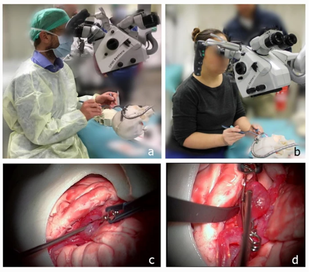
시뮬레이터 및 스터디 참여 그림 표현: 시뮬레이터에서 동맥류 모델을 조작하고 클리핑을 시도하는 전문 신경혈관 외과의사. b 젊은 레지던트 신경외과 클리핑. c 클리핑 시도. c 클리핑 후
신경외과 대학원 교육에서 시뮬레이터의 역할에 대한 향후 검증 연구의 실현 가능성을 평가하기 위해, 전문 신경외과 의사가 참가자의 클리핑 성과를 평가하여 그룹 간 비교를 실시했다.
그룹 A 및 그룹 B의 결과는 아래 차트를 참조하십시오.
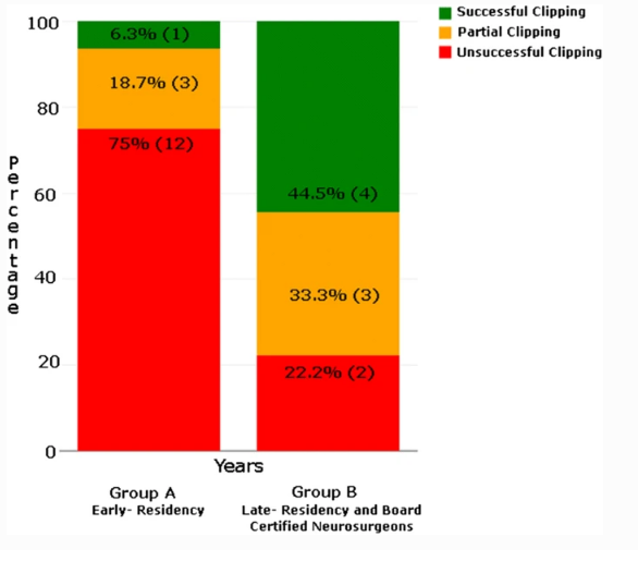
클리핑의 특성(성공, 성공 및 부분적으로 완료) vs 그룹 A와 그룹 B 참가자 사이의 다년간의 경험.
3D 프린팅은 항공우주, 건설, 에너지와 같은 다른 산업에도 상당한 이점을 제공하면서도 환자 맞춤형 치료를 제공할 수 있는 능력 때문에 특히 의학의 면모를 변화시키는 데 인상적이었다. 이 연구는 전통적인 교육 형태와 비교해서도 좋은 증거를 제시한다. 참가자의 80% 이상이 이 연구가 "이론적이고 전통적인 학습의 좋은 대안"이라고 생각했다.
그는 "현재까지 신경외과의사들은 환자의 결과라는 한 가지 성과 지표만을 사용해 왔다"고 말했다. 우리의 시뮬레이터는 기술을 향상시키고 유지하기 위한 대체 수단을 포함할 수 있을 것이다."라고 연구자들은 결론지었다.
"현재 시뮬레이터가 미래 신경외과의 훈련과 교육에서 품질보증을 측정, 평가, 유지하는데 귀중한 임상 도구임을 확인하기 위해서는 더 많은 연구가 필요하다."
No one wants to be operated on by a surgeon who has little hands-on training, but throughout history, finding suitable models or cadavers has been challenging for medical students who want nothing more than the luxury of many quiet hours perfecting their procedures as they continue on their way to being experienced doctors.
3D printing has brought a lot to the (operating) table for doctors worldwide, as well as to patients, their families, and medical students and doctors. 3D printed models and guides, whether displaying organs like the kidney, head and neck tumors, or hip fractures, allow for better diagnosis and treatment, as well as offering a comprehensive visual aid for everyone involved.
Simulation devices, while more complex, are also extremely helpful to students and doctors. Now, Swiss medical researchers from the University of Bern have developed a new training method, detailed in the recently published “Neurosurgical simulator for training aneurysm microsurgery—a user suitability study involving neurosurgeons and residents.”
Currently, training for intracranial aneurysms is limited because of the difficulty level involved in such microsurgery. Medical students and surgeons typically gain experience through watching and assisting during procedures, performing surgery under the watchful eye of skilled doctors, as well as studying videos. While there are some types of virtual reality simulation available, the researchers were motivated to improve on the technology with patient-specific models that even include features like blood circulation and pulsatility–something that is very important for training purposes but historically has been lacking.
Considering the delicate nature of intracranial aneurysm microsurgery, it is critical for students to understand the vascular nature of the aneurysm as well as exactly what happens in the case of a rupture. Students and doctors training should understand the surgical process overall, along with learning how to spot occlusions (blockage). It is concerning that because models and other experiential education are so hard to come by, there may just continue to be a lack of trained cerebrovascular neurosurgeons.
The goal of this study was to create a progressive simulator based on a 3D printed model that would be able to train both residents and neurosurgeons in clipping intracranial aneurysms. The materials used are meant to mimic human tissue, including:
-Arterial wall patency
-Thickness
-Elasticity
-Pulsatile blood flow
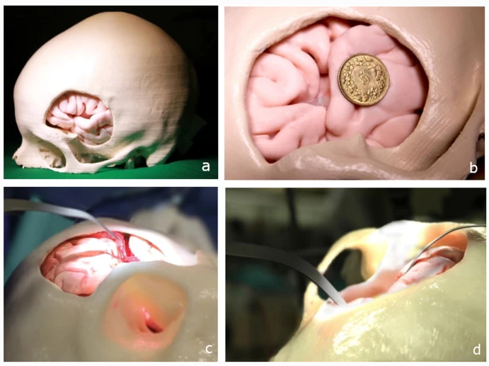
Representation of the model during the training study: a Patient-specific 3D-printed trephined skull with brain model. b MCA aneurysm model located in the left Sylvian fissure. c Pulsating blood vessel and access to the pathology of the model. d Brain retractor during manipulation by resident.
Twenty-five neurosurgeons and residents participated in the research study. Sixteen were in their early residencies with less than four years of training in the field of neurosurgery; nine were in late residencies and already board-certified, with anywhere from four to fifteen years of neurosurgical training. The residents and surgeons evaluated the efficacy of the new simulator, using a five-point scale to grade the following:
-Surgical simulation anatomy
-Realism
-Haptics
-Tactility
-General usage
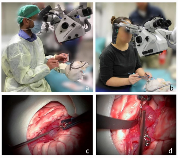
Pictorial representation from the simulator and study participation: a Expert neurovascular surgeon manipulating the aneurysm model in the simulator and trying to clip. b Young resident neurosurgeon clipping. c Attempt to clipping. d Exploration after clipping
In order to evaluate the feasibility of a future validation study on the role of the simulator in neurosurgical postgraduate training, an expert neurosurgeon assessed participants’ clipping performance, and a comparison between groups was done.
See the chart below for results from group A and group B.

Nature of clipping (successful, unsuccessful, and partially done) vs. years of experience between group A and group B participants.
3D printing, while offering substantial advantages to other industries like aerospace, construction, and energy, has been particularly impressive in changing the face of medicine due to the ability to offer more patient-specific treatment; this study offers good evidence of that in comparison to traditional types of training. Over 80 percent of the participants thought the study “was a good alternative to theoretical and conventional learning.”
“To date, neurosurgeons have been using only one indicator of performance: patient outcomes. Our simulator could incorporate alternative measures to improve and maintain skills,” concluded the researchers.
“Further studies are needed in order to confirm that the present simulator is a valuable clinical tool for measuring, evaluating, and maintaining quality assurance in the training and education of future generations of neurosurgeons.”
○ 출처 :


