[해외]미네소타 대학교 연구원들은 3D 바이오 프린팅을 사용하여 박동하는 인간의 심장을 만듭니다.
- 2020-07-09
- 관리자
○ 본문요약 :
미네소타 대학교 (University of Minnesota)의 연구원 들은 새로운 바이오 잉크를 개발하여 기능적인 3D 인쇄 박동 인간의 심장을 만들 수있게했습니다.
다 능성 줄기 세포를 사용하여 생산 된 세포가 함유 된 생체 재료를 통해 연구팀은 이전보다 더 많은 챔버, 심실 및 더 높은 세포벽 두께로 대동맥 복제물을 3D 인쇄 할 수있었습니다. 미래에,이 과정을 이용하여 복제 된 장기는 다양한 약물 및 장치에 대한 테스트 베드를 제공 할뿐만 아니라 유전자 질환에 대한 모델을 제공 할 수 있습니다.
Researchers from the University of Minnesota have developed a novel bio-ink, enabling them to create a functional 3D printed beating human heart.
The cell-laden biomaterial, produced using pluripotent stem cells, allowed the research team to 3D print an aortic replica with more chambers, ventricles and a higher cell wall thickness than was previously possible. In future, organs replicated utilizing this process, could provide a test bed for a variety of drugs and devices, as well as a model for genetic diseases.

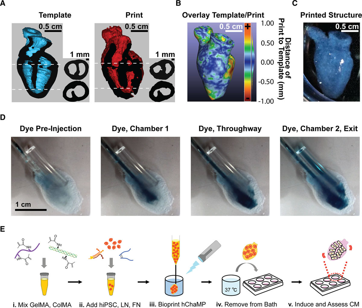
3D 프린트 하트의 정확성을 보여주는 제작 과정과 오버레이 (위)를 보여주는 다이어그램. 순환 연구 저널을 통한 이미지.
새로운 바이오 잉크를 생성하는 과정은 젤라틴 메타 크릴 레이트 (GelMA) 기본 재료로 시작되었으며, 이후 바이오 잉크를 인쇄 가능하게 만들기 위해 광 활성화를 통해 가교되었습니다. 줄기 세포 및 ECM 단백질뿐만 아니라 화학 물질 인 피브로넥틴 및 라미닌 -111을 추가하면 심근 세포 분화 및 구조적 완전성을지지하는 것으로 밝혀졌다. 이 제형은 0.1 mg DNA / g의 겔의 세포 밀도를 달성하였으며, 이는 천연 심장 조직과 동일한 크기로 실험에 이상적이다.
3D 팀은 인간의 심장과 새로 고안된 바이오 잉크의 자기 공명 영상 스캔을 사용하여 2 개의 챔버와 용기 입구 및 출구가있는 줄기 세포가 담긴 구조물을 인쇄했습니다. 세포가 충분한 밀도로 곱한 후, 연구진은 세포를 구조 내에서 분화시켜 심장 내에서 다른 역할을하게했습니다. 심실 사이의 중격은 부분적으로 제거되어 영양 전달을위한 입구를 제공하고, 구조는 2 개의 혈관 연결로 제한되었지만, 심장의 성능은 탄력적이고 기능적인 것으로 입증되었다.
The process of creating the new bio-ink began with a gelatin methacrylate (GelMA) base material, which was later cross-linked via photoactivation to make the bio-ink printable. Further adding the chemicals fibronectin and laminin-111, as well as stem cell and ECM proteins, was found to support cardiomyocyte differentiation, and structural integrity. This formulation achieved a cell density of 0.1 mg DNA/g of gel, which is on the same order of magnitude as native cardiac tissue, making it ideal for experimentation.
Using a magnetic resonance imaging scan of a human heart and the newly devised bio-ink, the team 3D printed stem cell–laden structures with two chambers and a vessel inlet and outlet. After the cells had multiplied to a sufficient density, the team differentiated the cells within the structure, giving them different roles within the heart. While the septum between the ventricles was partially removed to provide entry for nutrient delivery, and the structure was limited to two vascular connections, the heart’s performance proved resilient and functional.
○ 출처 : 


