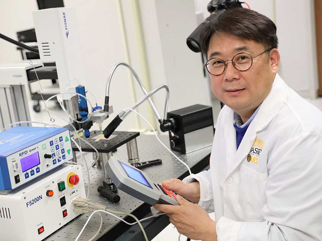
[해외]VIRGINIA COMMONWEALTH UNIVERSITY 교수, 3D 프린팅을 사용하여 암 연구 발전
- 2020-05-17
- 관리자
○ 본문요약 :
버지니아 커먼 웰스 대학 (VCU) 의 인문 과학부 조교수 는 3D 프린팅을 사용하여 종양 세포의 라이브 모델을 만들어 암 연구자들이 질병의 진행 상황을 더 잘 이해할 수있게했습니다.
An assistant professor from the Virginia Commonwealth University (VCU) College of Humanities and Sciences has used 3D printing to create live models of tumor cells, which could enable cancer researchers to better understand the disease’s progression.

암 연구에서 2D 프린팅에서 진화
연구원 유럽 분자 생물학 연구소 독일은 2 차원 구조를 정확하게 기본 종양 미세 환경의 특성을 복제 지장이 있다고 12 월 2018 그들의 연구에서 발견. 3D 세포 배양은 실제 조직의 특이성을 더 잘 모방하고, 더 빠른 속도로 수행하며,보다 복잡한 행동을 나타내는 세포 간 상호 작용을 확립하는 것으로 밝혀졌다.
Evolving from 2D printing in cancer research
Researchers from The European Molecular Biology Laboratory in Germany found in their study in December 2018, that 2D structures have difficulty in accurately replicating the characteristics of native tumor microenvironments. 3D cell cultures were found to establish cell-to-cell interactions that could better mimic the specificity of real tissues, doing so at greater speed, and displaying more complex behaviors.
암 퇴치의 3D프린팅 방법
다른 교육 기관의 과학자들과 연구원들은 암 종양을 더 잘 이해하고 치료하기 위해 3D 프린팅을 사용하여 라이브 3D 세포 모델을 만들었습니다.
2020 년 4 월 미국과 독일의 연구원들은 공격적인 뇌종양 인 교 모세포종 (GBM) 을 연구 하는 새로운 방법을 제시 했습니다. 그들은 장기 배양 및 약물 전달을 허용하기 위해 관류 혈관 채널을 특징으로하는 인간 뇌 세포 및 생체 물질의 모음을 사용했다. 조직 구조물의 비 침습적 평가를 위해 3D 영상 기술을 요약하여 사용 하였다.
3D printing methods of combating cancer
Scientists and researchers from other academic institutions have also used 3D printing to create live 3D cell models, with the aim of better understanding and treating cancerous tumors.
In April 2020 researchers from the USA and Germany presented a new way of studying glioblastoma (GBM), an aggressive type of brain cancer. They used a collection of human brain cells and biomaterials featuring perfused vascular channels, to allow long-term culture and drug delivery. 3D imaging technology was summarily used for the noninvasive assessment of the tissue constructs.
○ 출처 : 
https://bit.ly/3cJtzQA


